03 Education · Interactive 3D
Vertebrate Lab 3D
An interactive 3D anatomy and virtual dissection lab for high school science classrooms.
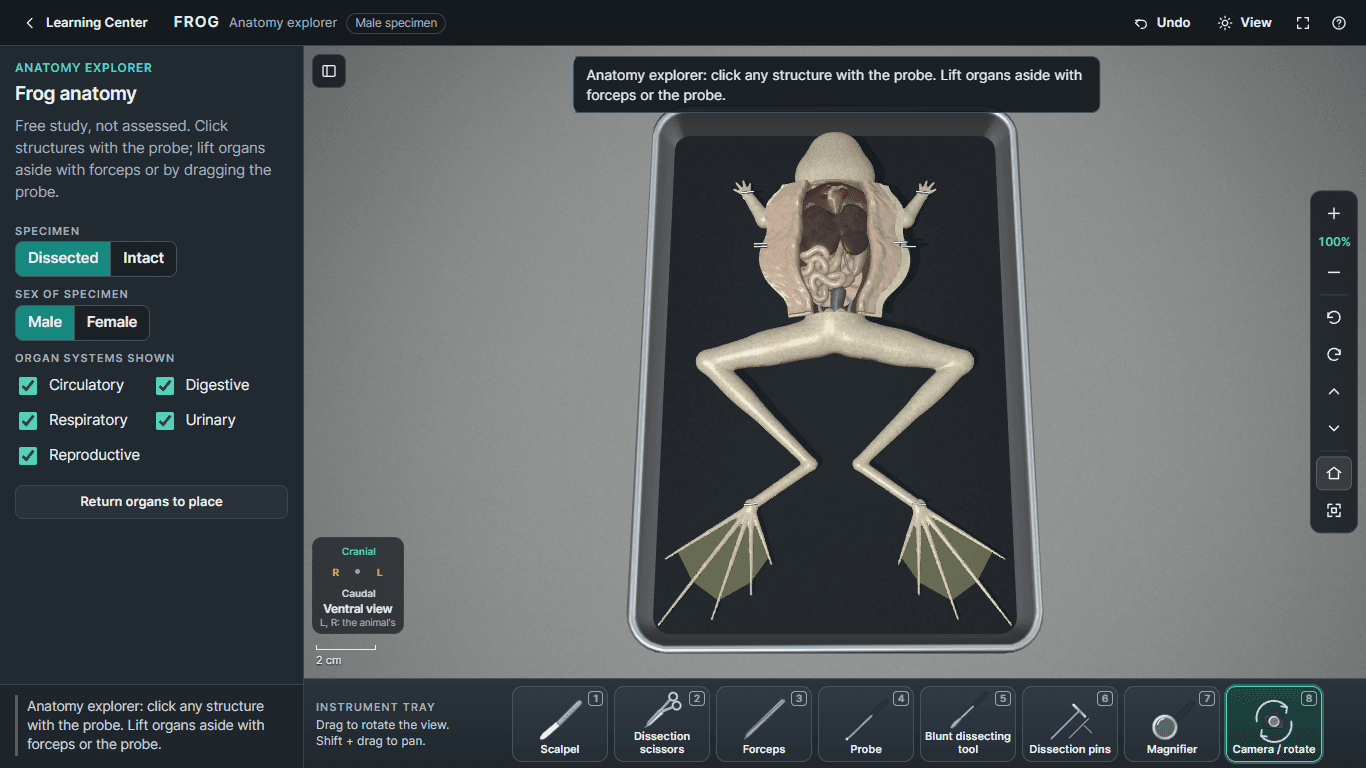
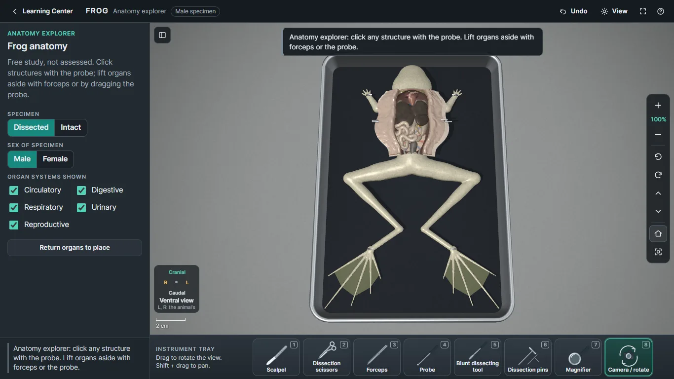
The frog anatomy explorer with organ systems shown.
Overview
Dissection is one of the most memorable parts of a science class. It is also hard to prepare for, and specimens are limited. Vertebrate Lab 3D gives students a place to practice first. They can learn where structures are, how the instruments work and what each step of a dissection involves before they ever pick up a real scalpel.
It was built for high school agriculture, animal science and biology classrooms, with the Career and Technical Education Academy in Sioux Falls in mind. It runs on school Chromebooks with just the built-in trackpad, and it works without an internet connection.
Vertebrate Lab 3D is an independent project. It is not an official product of any school or district.
What it does today
Features
Five specimens in 3D
Frog, rat and cat for small animal science; a fetal pig and an isolated sheep brain for large animal science. Male and female specimens where it matters.
Virtual instruments
Scalpel, dissection scissors, forceps, probe, blunt dissecting tool, pins and magnifier, all usable with a Chromebook trackpad. No external mouse needed.
Guided and independent labs
Step-by-step guided dissections with hints and on-model "Show Me" markers, and independent labs where students work on their own.
Anatomy explorer
Free study mode for clicking structures, lifting organs aside and toggling organ systems to see how they fit together.
Real specimen comparisons
Many structures link to genuine specimen photographs with source and rights information, so students can compare the model to the real thing.
Student reports and saved progress
Lab progress is saved on the device and students can download a report of their work.
Works offline
The whole application is one HTML file. No installation, login, account, subscription or server is required.
Real screenshots
Screenshots
The frog anatomy explorer with organ systems shown. 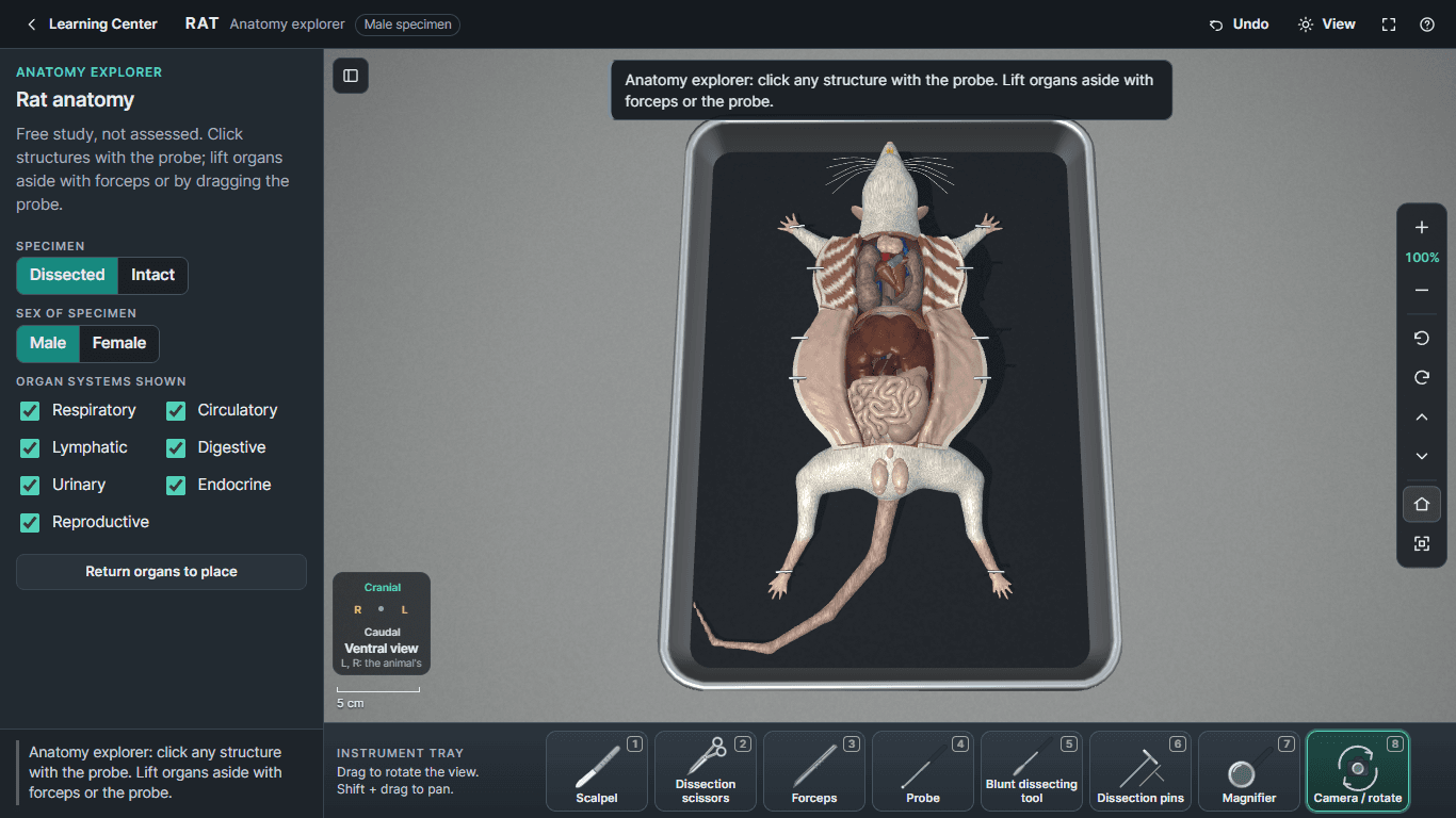
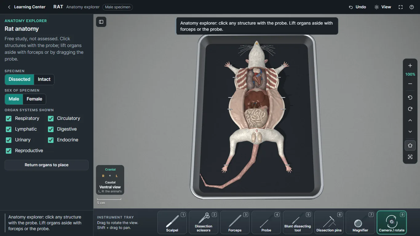
Rat anatomy, ventral view. 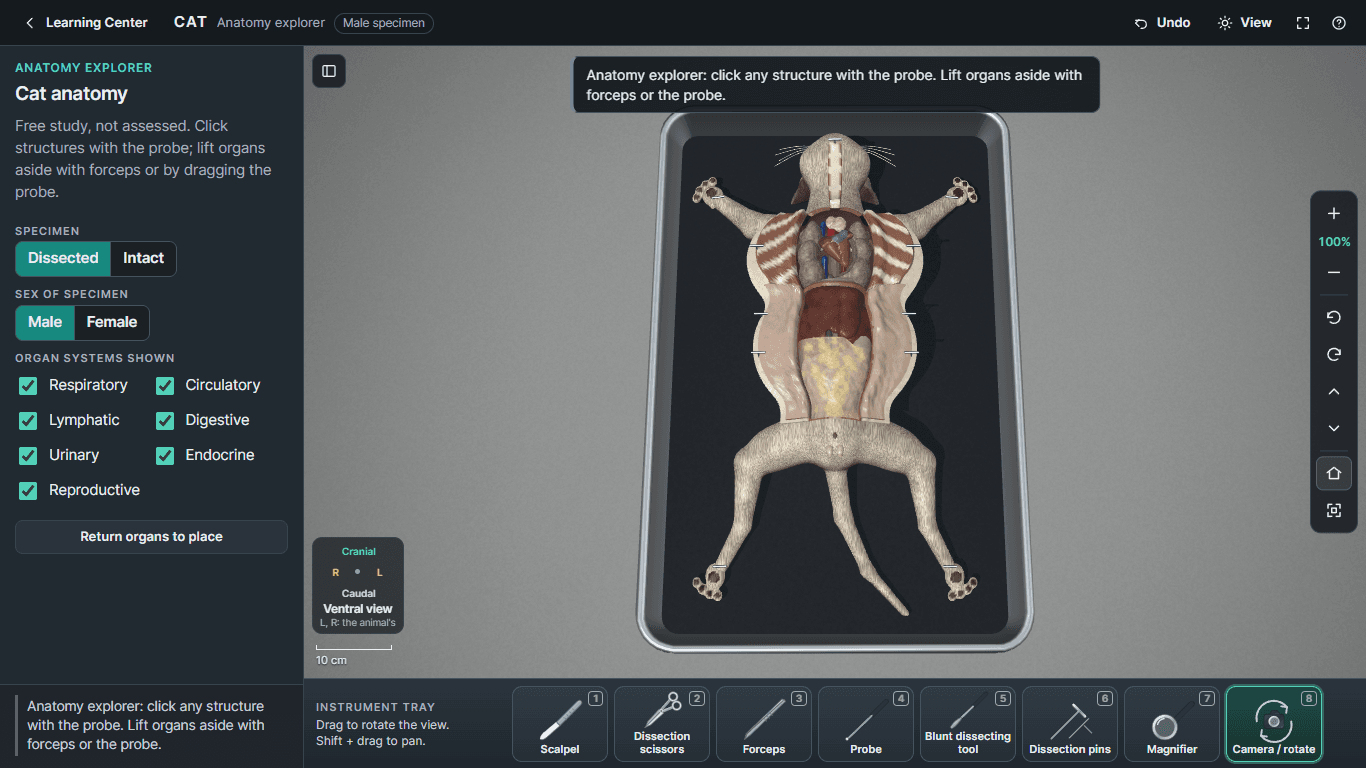
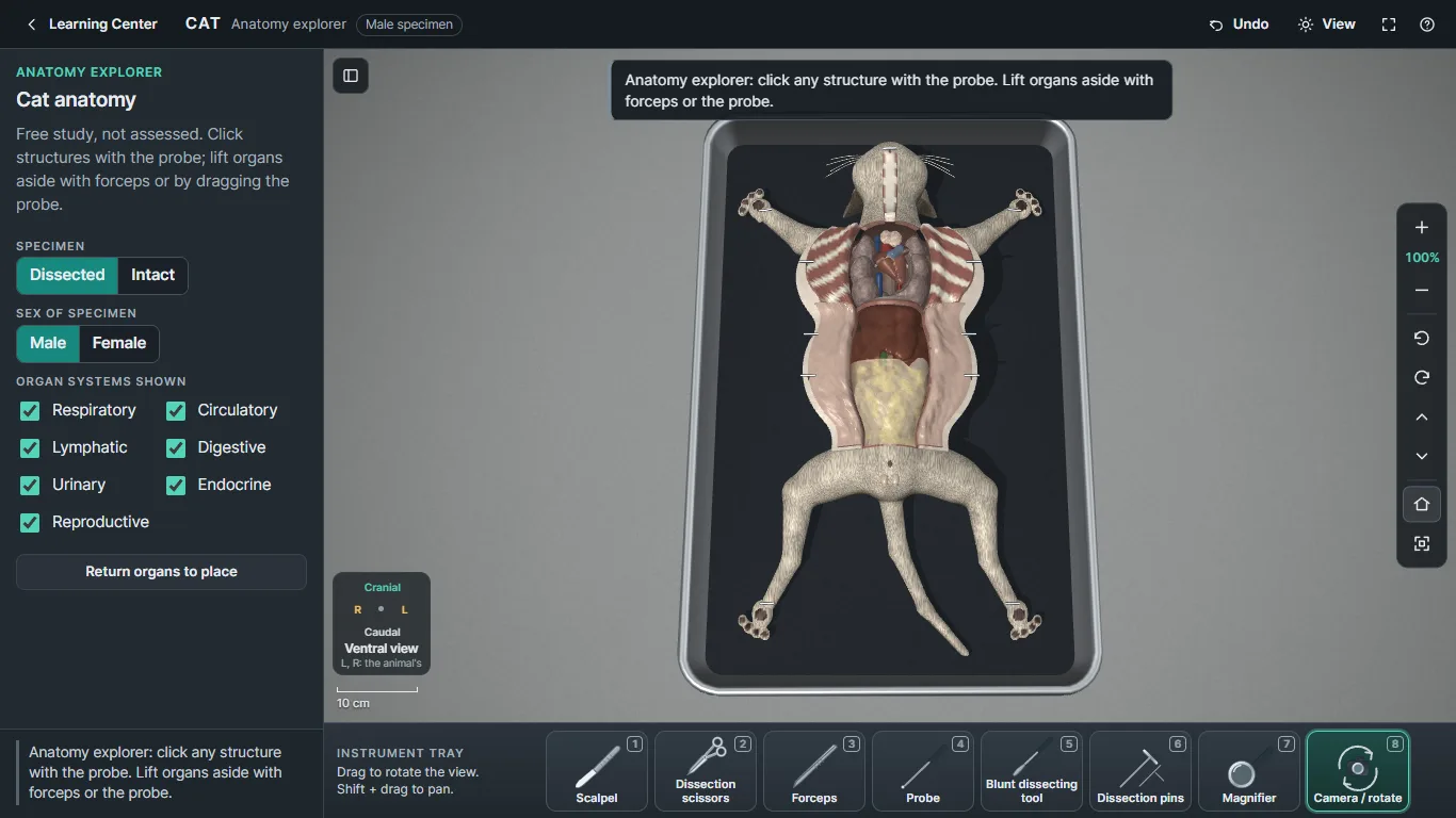
Cat anatomy, ventral view. 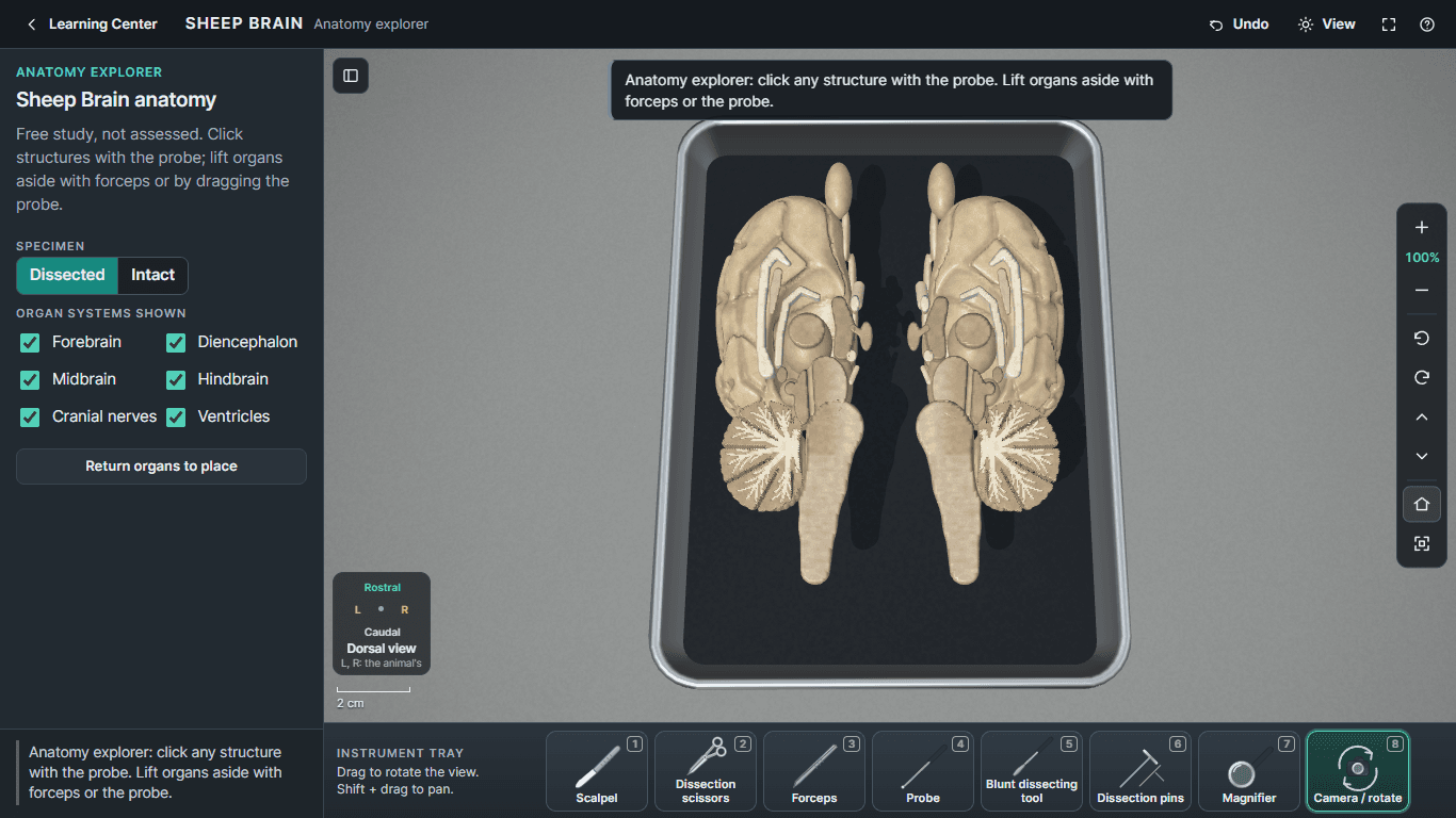
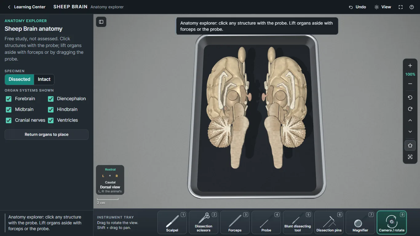
The sheep brain, sectioned along the midline.
Development
Current status
Status: Release candidateRelease candidate. Automated testing passes; teacher and anatomy-expert review and validation on classroom Chromebooks are still in progress.
Built with
- JavaScript
- WebGL
- Single-file HTML
Planned next
- Teacher and anatomy-expert content review
- Validation on classroom Chromebooks and display boards
- Broader real-specimen photograph coverage
Good to know
- Virtual dissection is a preparation and learning aid; it is not a replacement for teacher instruction.
- Real specimen photographs cover most, but not all, structures.